Rhea Medical Center’s Radiologists
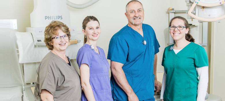
The experienced radiologist staff at Rhea Medical Center are Dr. Roger Miller and Dr. Lebron Lackey of Cleveland Radiology Associates. One of the two radiologists is at the hospital five days a week. Their expertise is an important asset to Rhea Medical Center.
General Radiology
Rhea Medical Center offers a wide variety of general radiology examinations, including examinations of the chest, head, abdomen, spine, and extremities. Most general radiology examinations do not require an appointment; however, an order from your doctor is required for all examinations.
A scheduled appointment is required for certain series, such as CT, MRI, Ultrasound, Mammography, upper gastrointestinal, barium enemas, barium swallowing tests, and intravenous pyleograms, etc.
Mammography
What is a mammogram?
A mammogram is an X-ray from the side and top of the breast. Mammograms are an important tool in early detection of breast cancer because they can detect a lump three to five years before your doctor can feel it. During a mammogram, your breasts are pressed between two pieces of plastic for a few seconds, while a minimal X-ray dose, similar to that of a dental X-ray, is applied.
Why might I need a mammogram?
All women share some risk of breast cancer. In the United States it is estimated that there will be nearly 213,000 new cases of female invasive breast cancer this year. Experts predict almost 41,000 deaths from the disease. But the good news is that the number of women who obtain breast mammograms has doubled since National Breast Cancer Awareness Month began over twenty years ago, and now the death rates from breast cancer have declined, mostly due to earlier detection and improved treatment. Mammography screenings are a woman’s best chance for detecting breast cancer early, according to the American Cancer Society:
- Women 40 years and older should get a mammogram every 1 to 2 years.
- Women who have had breast cancer or other breast problems or who have a family history of breast cancer, might need to start getting mammograms before age 40 or they might need to get them more often. Talk to your doctor about when to start and how often you should have a mammogram.
Mammography at Rhea Medical Center
Rhea Medical Center has the most advanced Digital Mammography technology available.
Lead Mammographer Edie Shaver has worked hard to make getting a mammogram as pleasant an experience as possible for her patients. Women who get their mammograms at Rhea Medical Center enjoy a warm and comfortable environment, and benefit from the positive and friendly attitude of Shaver and the rest of the mammography staff. In addition to regular office hours, Rhea Medical Center offers after-hours appointments for women wishing to get a mammogram without taking time off from work. The evening schedule is available Mondays, Tuesdays and Wednesdays.
Women’s Waiting Area
What to Expect:
Preparation: Going in for your first mammogram may seem a little intimidating at first, but Shaver says that the mammography staff will counsel women on any concerns they may have about a screening or its results prior to the mammogram. Patients can be confident knowing that the facilities and equipment at Rhea Medical Center are as good as any in the region.
Procedure: During the mammogram, your breasts will be pressed between two pieces of plastic for a few seconds, which may be briefly uncomfortable. “Some people think that mammograms are painful, but with the new equipment, they really aren’t,” said Shaver. “With this machine, the whole process is quick and easy.”
Learn More
Articles and Links about Breast CancerWomen’s Health Newsletter
Common Brast Cancer Facts, Stats, and Myths
Breast Cancer Awareness Month focuses on Early Detection
Ultrasound
What is an ultrasound?
During an ultrasound exam, (also known as a sonogram), sound waves far above the range of human hearing penetrate your body. When your internal organs reflect back the sound waves, a computer records and interprets the resulting echoes and generates an image of the area of your body being examined. An ultrasound does not use X-rays; therefore, it is appropriate even for pregnant women, who are advised to avoid X-ray and CT scans.
Advanced Ultrasound Technology
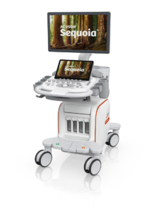
 Rhea Medical Center is proud to introduce our NEW Sequoia Ultrasound System, the most advanced ultrasound imaging technology available in our region! This state-of-the-art system boasts a unique design that allows for focus on multiple fields of view at all depths, along with speed of sound correction technology to provide improved resolution, and vascular capabilities to improve detection of vessels and resolution within and between organs.
The addition of the Sequoia is just one more way Rhea Medical Center continues to expand and grow our services as we continue to lead the way in healthcare excellence for our community!
Ultrasound at Rhea Medical Center
Because of its advanced equipment, Rhea Medical Center offers a wide range of ultrasound procedures, including:
- Abdomen
- Breast
- Obstetric/Fetal
- Pelvis
- Renal (kidneys)
- Testicular
- Thyroid
- Vascular
- Echocardiography
- Stress Echocardiography
Equipment: Rhea Medical Center was one of the first hospitals in southeast Tennessee to acquire the Acuson Sequoia.
Our Ultrasound system is the most advanced system available on the market. This equipment is used mainly by large metropolitan teaching hospitals. It provides clearer images of the body and allows us to see abnormalities at a smaller, earlier stage. The Sequoia allows us to complete exams that lesser ultrasound equipment would not be capable of performing.
What to Expect
The Procedure: An ultrasound technologist will spread a warm transmitting gel over the area of your body to be scanned and then run a wand-like instrument, (called a transducer) lightly through the gel. A video screen will display a moving image of the area examined, and the image will be photographed for analysis.
Bone Mineral Density
What is a Bone Mineral Density test?
A Bone Mineral Density test, also referred to as bone densitometry, uses special X-rays to measure how many grams of calcium and other bone minerals are packed into a segment of bone. The higher your mineral content, the denser your bones. BMD is an important diagnostic tool that not only measures the amount of calcium in certain bones, but also can be used to estimate the risk of a fracture. The test is easy, fast, painless, and non-invasive. Doctors use a bone density test to determine if you have, or at risk of, osteoporosis.
What is osteoporosis?
Osteoporosis is a disease in which bones become fragile and more likely to break. If not prevented or if left untreated, osteoporosis can progress painlessly until a bone breaks. These broken bones, also known as fractures, occur typically in the hip, spine, and wrist. Millions of Americans are at risk of getting osteoporosis.
Why might I need a Bone Mineral Density Test?
Bone Mineral Density tests are particularly important for women, who are at higher risk of getting osteoporosis. The test should be considered when:
- An X-ray reveals low bone mass or possible osteoporosis
- Menopause occurs prior to age 45 and the patient is not taking estrogen
- A woman is age 65 or older
- A post-menopausal woman sustains a fracture
- There is a family history of osteoporosis
- Steroids have been (or are) taken regularly
- Hyperthyroidism, diabetes, liver/kidney disease, or rheumatoid arthritis is present
The older you get, the higher your risk of osteoporosis because your bones become weaker as you age. You are also at greater risk for osteoporosis if you’re white or of Southeast Asian descent. Other risk factors include low body weight, personal history of fractures, and using certain medications that can cause bone loss.
What to Expect
Preparation: Dual Energy X-ray Absorptiometry or DEXA is the most common method used to measure bone density and requires no patient preparation.
Procedure: The patient simply lies on a padded table during the scan of a particular part of the body such as the lower spine and hip. The test period is short, usually only several minutes. A radiologist reads and compares the results to normal values and prepares a concise report for the referring physician.
Learn More:
Articles and Links about Osteoporosis and Bone Health
CT (Computer Tomography) Scan
What is a CT Scan?
A CT scan is a procedure that generates a series of computerized images that can be used to detect conditions that often do not show up on conventional X-ray images. The fine detail of a CT scan shows a clear picture of soft tissues, internal organs, and bone structures, including: the brain, chest, abdomen, pelvis, and spine.
CT at Rhea Medical Center
Rhea Medical Center has a 16-slice CT scanner. This CT scanner improves both the speed at which images can be obtained (a chest scan takes just 10-15 seconds) and the quality of images produced. The three-dimensional visualizations of internal organs and structures produced by the CT scanner provide Rhea Medical Center radiologists with the material they need to make accurate readings.
What to Expect
Preparation: Some exams require no special preparation, but in other cases we may ask you to fast for four hours before your test. If you’ve been scheduled for a CT scan of the abdomen or pelvis, you’ll need to drink a barium mixture at home before your exam. If you are scheduled for a CT scan of the head or neck, we’ll ask you to remove any objects, such as hairpins, jewelry, hearing aids, dentures, or glasses, which might interfere with the X-rays.
Procedure: During the procedure, you will lie down on a table, which will be positioned in the CT scanner. You may hear noises as the machine takes its images. You will be asked to lie still. The test is painless. It’s best just to relax and rest while it is being performed. Because of Rhea Medical Center’s state-of-the-art equipment, the test will only take a few minutes at the most.
Nuclear Medicine
Nuclear Medicine is a medical specialty that uses safe, painless, and cost-effective techniques to document organ function and structure. An integral part of patient care, Nuclear Medicine is used in the diagnosis and management of diseases. Nuclear Medicine uses a very small amount of radioactive materials or radio pharmaceuticals to diagnose and treat disease. Radiopharmaceuticals are substances that are attached to specific organs, bones, or tissues. The radiopharmaceuticals emit gamma rays that can be detected externally by special types of cameras: gamma or PET cameras. These cameras work in conjunction with computers used to form images that provide data and information about the area of the body being imaged.
MRI
What is MRI?
Magnetic Resonance Imaging is a technique that enables physicians to see internal organs without using surgery or X-rays. MRI does not use radiation like traditional X-ray modalities. A sophisticated computer enhances images created by a magnetic field and radiofrequency waves. These images are transformed into cross-sectional views of the organ or area being studied. In some cases, the injection of a contrast agent may be needed to enhance the detail of particular body parts.
Why might I need an MRI Scan?
MRI is very useful in diagnosing a variety of conditions and disorders affecting:
- The central nervous system: the soft tissue parts of the brain and spinal cord.
- Orthopedic structures: internal bone architecture and joints, such as the knee, shoulder, jaw, wrist, and ankles. MRI is also the best imaging technique for cartilage, muscle, and ligaments.
- Abdominal and pelvic organs: the pancreas, liver, adrenal glands, and reproductive organs
- Blood vessels: arteries and veins
What to Expect
Preparation: Usually there are no special preparations required for an MRI scan. You can continue to take medications as usual. Metal objects may interfere with the magnetic field of an MRI scanner. It is very important that we know about any metallic devices that you may have in your body. These devices may prevent you from having an MRI scan. Please let us know if you have any of the following:
- Cardiac pacemaker
- Internal electronic device
- Heart valve
- Coronary artery stent
- Metal surgical clips or aneurysm clips
- Hearing aid or implants
- Shunts
- Artificial joints/ metal rods
- Embedded shrapnel
- Metal in eyes (which may have resulted from sheet metal work)
All metal objects must be left outside the examination room. Such objects include:
- Jewelry and watches
- Hairpins
- Dentures
- Credit Cards
If you are wearing any clothing containing metal, such as zippers, snaps, underwire bras or bra hooks, you will be asked to undress and put on an exam gown.
Procedure: You will lie on a table that positions you within the MRI unit, a large open-ended tube that will surround your body while you are being scanned. A trained and licensed radiologic technologist will observe you from another room, talking with you via intercom, while he or she operates the computer that controls the MRI unit. You will hear some tapping noises, as the computer generates the MRI images; during this time you should lie motionless so that the images are as clear as possible. If you feel uncomfortable, please tell the technologist, who can hear you at all times. We will provide you with earplugs or headphones.
